X-rays films are photographic representations in black and white of bones and tissues of the human bodies. Examining x-rays helps doctors to have an internal view of the patient's body which supports the external signs and symptoms observed by the patient. Signs and symptoms of different diseases appear to be the same hence the use of X-rays to differentiate the diseases and perfectly diagnose a patient.
The stated signs and symptoms suggest that the patient is suffering from a chest problem and to specifically identify the disease, the results show a clot in the lungs which makes us conclude that the patient is suffering from Bacterial pneumonia since it's the only disease that can be transmitted from one person to another (Brooks& Abdallah, 2020). Other diseases that the patient could suffer from include; Pulmonary embolism, Influenza and Subacute bacterial endocarditis. Bacterial pneumonia out ruled all other diagnoses due to the inflammation of the lungs.
The patient's history describes a man who indulged in drugs and unprotected sex with the past four cases of drug abuse and turning positive for sexually transmitted diseases such as Hepatitis C, Thrush. Bacterial pneumonia mostly affects people who used to smoke and given the fact that the patient is living in a group home, they must be recovering from the abuse of tobacco. The medical history of the patient helped us realize past or current medical conditions they suffered from and how they relate to the current condition for us to get a perfect diagnosis. Testing HIV/AIDS positive indicated that the patient had a weak immune system which could not prevent the transmission of diseases from person to person. On the other hand, physical examination enabled us to know the age of the patient to identify several conditions affecting their age bracket.
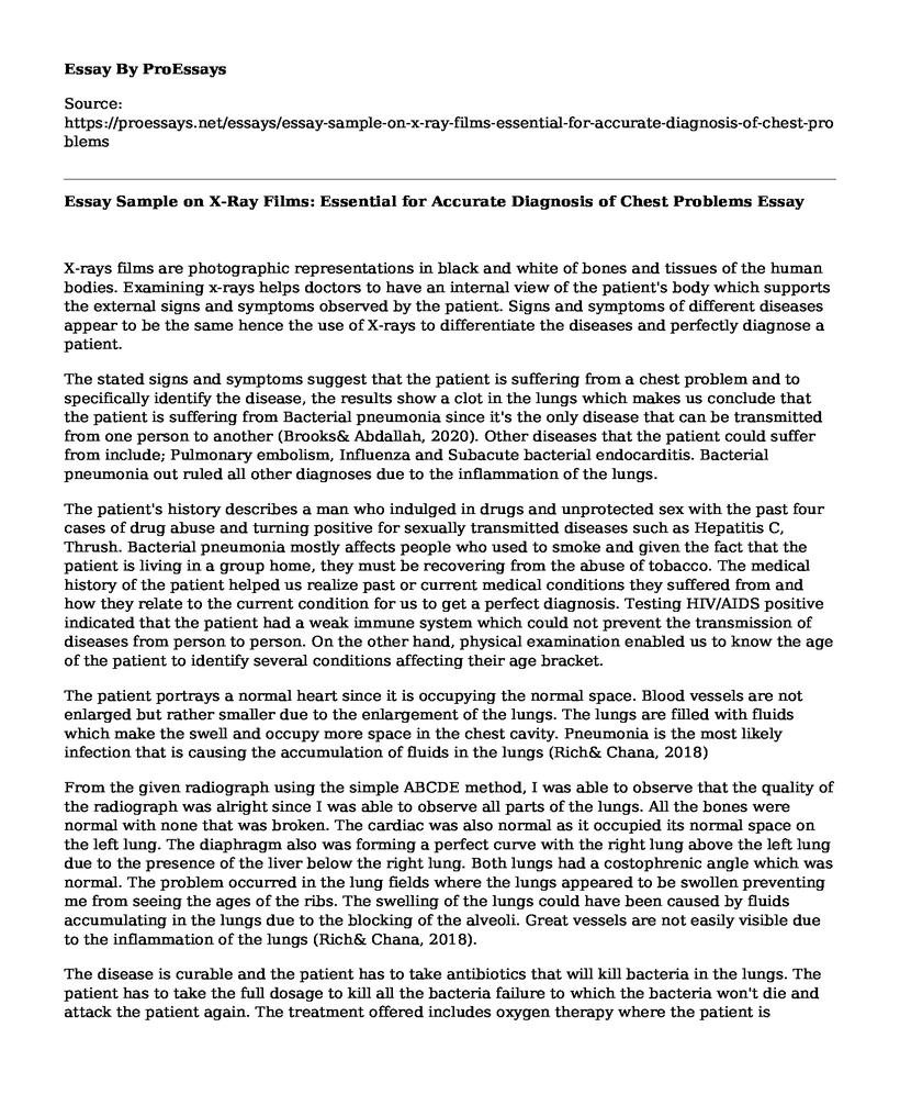
The patient portrays a normal heart since it is occupying the normal space. Blood vessels are not enlarged but rather smaller due to the enlargement of the lungs. The lungs are filled with fluids which make the swell and occupy more space in the chest cavity. Pneumonia is the most likely infection that is causing the accumulation of fluids in the lungs (Rich& Chana, 2018)
From the given radiograph using the simple ABCDE method, I was able to observe that the quality of the radiograph was alright since I was able to observe all parts of the lungs. All the bones were normal with none that was broken. The cardiac was also normal as it occupied its normal space on the left lung. The diaphragm also was forming a perfect curve with the right lung above the left lung due to the presence of the liver below the right lung. Both lungs had a costophrenic angle which was normal. The problem occurred in the lung fields where the lungs appeared to be swollen preventing me from seeing the ages of the ribs. The swelling of the lungs could have been caused by fluids accumulating in the lungs due to the blocking of the alveoli. Great vessels are not easily visible due to the inflammation of the lungs (Rich& Chana, 2018).
The disease is curable and the patient has to take antibiotics that will kill bacteria in the lungs. The patient has to take the full dosage to kill all the bacteria failure to which the bacteria won't die and attack the patient again. The treatment offered includes oxygen therapy where the patient is equipped with skills on how to breathe in their current condition. 100 grams of omadacycline are taken every 12 hours and an additional 100 grams after every 24 hours for four weeks due to the weak immune system of our patient( Liu et al., 2018) To ease the chest pains, the patient should take 200 grams of panadol painkiller after every 8 hours for five days. The patient should also take lots of fruits and vegetables. The patient should also avoid crowded places to contain the spread and have enough rest. The CT-scan is the other test I would do to the patient to have a closer look at the lungs and assess the damage caused by the infection (Stets et al, 2019).
References
Brooks, W. A. (2020). Bacterial Pneumonia. In Hunter's Tropical Medicine and Emerging Infectious Diseases (pp. 446-453). Content Repository Only!.Liu, S., Paul, P., Su, J., & Xu, C. (2018). U.S. Patent No. 9,952,965. Washington, DC: U.S. Patent and Trademark Office.
Rich, C. (2018). Radiology, Education, Community EM. Radiology.Stets, R., Popescu, M., Gonong, J. R., Mitha, I., Nseir, W., Madej, A., ... & Manley, A. (2019). Omadacycline for community-acquired bacterial pneumonia. New England Journal of Medicine, 380(6), 517-527.
Cite this page
Essay Sample on X-Ray Films: Essential for Accurate Diagnosis of Chest Problems. (2023, May 02). Retrieved from https://proessays.net/essays/essay-sample-on-x-ray-films-essential-for-accurate-diagnosis-of-chest-problems
If you are the original author of this essay and no longer wish to have it published on the ProEssays website, please click below to request its removal:
- Paper Example on Skin Pressure Injuries
- Introducing Electronic Health Records to Nurses - Paper Example
- Adherence in Diabetes Patients Assignments Paper Example
- Research Paper on Background of the Gap in Nursing Education
- Endocrine Disruptors: Atrazine Paper Example
- Concept-Based Nursing Curriculum Paper Example
- Essay Example on Informatics Programs to Curb Spread of Infectious Diseases


