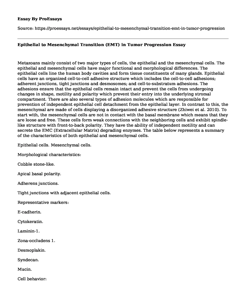
Metazoans mainly consist of two major types of cells, the epithelial and the mesenchymal cells. The epithelial and mesenchymal cells have major functional and morphological differences. The epithelial cells line the human body cavities and form tissue constituents of many glands. Epithelial cells have an organized cell-to-cell adhesive structure which includes the cell-to-cell adhesions; adherent junctions, tight junctions and desmosomes; and cell-to-substratum adhesions. The adhesions ensure that the epithelial cells remain intact and prevent the cells from undergoing changes in shape, motility and polarity which prevent their entry into the underlying stromal compartment. There are also several types of adhesion molecules which are responsible for prevention of independent epithelial cell detachment from the epithelial layer. In contrast to this, the mesenchymal are made of cells displaying a disorganized adhesive structure (Zhiwei et al. 2010). To start with, the mesenchymal cells are not in contact with the basal membrane which means that they are loose and free. These cells form weak connections with the neighboring cells and exhibit spindle-like structure with front-to-back polarity. They have the ability of independent motility and can secrete the EMC (Extracellular Matrix) degrading enzymes. The table below represents a summary of the characteristics of both epithelial and mesenchymal cells.

Epithelial cells. Mesenchymal cells.
Morphological characteristics:
Cobble stone-like.
Apical basal polarity.
Adherens junctions.
Tight junctions with adjacent epithelial cells.
Representative markers:
E-cadherin.
Cytokeratin.
Laminin-1.
Zona-occludens 1.
Desmoplakin.
Syndecan.
Mucin.
Cell behavior:
Non-motile Morphological traits:
Spindle-like.
Front-back polarity.
Attach to the extracellular matrix Representative markers:
Vimentin.
Fibroblast specic protein 1 (FSP1).
Snail.
Slug.
ZEB2.
Twist.
FOXC2.
Fibronectin.
Alpha smooth muscle actin.
N-cadherin.
Cell behavior:
Motile.
A change of any cell from the epithelial tissue to a mesenchymal cell is called the Epithelial-Mesenchymal Transition (EMT) and a change of a mesenchymal cell to an epithelial cell is referred to as the Mesenchymal-Epithelial Transition (MET) (Raghu et al., 2009). EMT is accompanied by complex biochemical events of both physical and cellular changes, which makes the epithelial cells lose their characteristics and acquire the mesenchymal traits. This process is particularly important in implantation, embryo formation, fibrosis, organ development, healing wounds and tissue regeneration. Apart from the above mentioned roles of EMT, it has been recently discovered that this process is a major part of epithelial cancer cell distribution throughout the body which is our concept of interest in this review. The plasticity of epithelial cells is essential for tumor progression. In the early stages of cancerous tumor development, the epithelial tumor cell undergo changes by commanding the EMT process to occur. This process has been one of the hottest fields in science recently and though faced with opposing controversies, the concept of tumor cells undergoing EMT has been demonstrated, in vitro, to be true by several scientific groups. To prove the existence of EMT in cancer had previously proven difficult because the mesenchymal cells formed from the tumor epithelial cells are indistinguishable from the normal surrounding fibroblasts but recently, a new method called the cell fate mapping has provided proof of EMT in cancer (Raghu et al., 2009). The primary tumors undergo EMT and through metastasis-a process combining local invasion, extravasation, intravasation, transport and colonization by cancer cells- the cells are spread from the site of the primary tumor and form a secondary tumor at a different site or in/on another organ. Through EMT, the cells undergo several changes which include; acquisition of invasive properties, down regulation of E-cadherin which is responsible for cell-to-cell adhesion, up regulation of plastic mesenchymal proteins such as vementin, smooth muscle protein actin and N-cadherin and deregulation of the Wnt pathway.
Before the understanding of the mechanism used by cancer cells to move from the primary tumor and develop in another locality, it was difficult to design a treatment strategy to stop the progression. However, with the connection between EMT and cancer, new therapeutics to target certain cancer EMT activities are being developed. For example, the twist1-mediated signaling pathways have been established as a target in cancer therapeutics. This pathway is believed to be rendered inactive by RNAi interference which promotes cellular senescence and growth halt which may control Twist1 mediated tumor metamorphosis. When the signaling pathway is interrupted, the EMT process is inactivated. This approach works through reducing cell migration and local invasion and increasing the rate of cancer cell apoptosis and is particularly promising in cisplatin-resistant lung cancer and breast cancer. Another importance of EMT in cancer is that it enables tumor progression i.e. the spread of tumors in the same locality or to a different part or organ in the body. For the tumor to spread, the primary tumor cells undergo EMT in order to change to mesenchymal cells which have motile and invasive properties. The mesenchymal traits enables the cells to move from the primary location to another locality. Once they get to the target point, this cells need to change back to epithelial cells in order to divide and grow into tumors (Honor et al., 2007). This is made possible by MET where the cells regain the epithelial traits. Apart from that, in the normal body functions, EMT is very important in; organ development, for example, in embryos where epithelial cells undergo EMT and migrate to a region where an organ needs to be formed and then undergo MET to form epithelial cells for the organ development to start; in damaged tissue and organ repair where surrounding cells undergo EMT to enable the process of new cell and tissue formation and in several other processes mentioned above. Several EMT-TFs in conjunction with other factors such as signaling and growth factors facilitate this process by down regulating E-cadherin and other junctional proteins.
Signaling pathways, growth factors and transcription factors that drive EMT
It is believed that the processes that govern EMT/MET are stimulated and regulated by many different stimuli, signal transduction pathways and transcriptional factors.
Signaling pathways
The full spectrum of signaling agents that activate EMT in tumor progression is not fully known. The genetic and epigenetic alteration is one of the factors thought to make the cells within the tumor respond to EMT-inducing signals from within the tumor. Some of the EMT inducing signals for many tumor progressions include HGF, EGF, FGF, PDGF, and TGF-b and appear to be the once responsible for initiating a series of EMT-inducing transcriptional factors. For the EMT process to occur successfully, it depends on the combined efforts of the following signal inducing proteins; MAPK, ERK, LEF, P13K, Akt, Smads, RhoB, b-catenin, Ras and c-Fos and the following cell surface proteins; aSb1 integrin and aVb6 integrin. (Honor et al., 2007)
TGF-b plays an important role as one of the most common signaling pathways in most tumors. As a growth factor, TGF-b is involved in prevention of tumor regeneration through suppressing epithelial cell proliferation. It is a member of the b family growth factors. However, it is not yet fully known how it causes tumor regeneration and consequently tumor progression but according to in vitro studies done recently, it can induce an EMT in certain type of cancer cells through one of its two signaling pathways. The conversion of TGF-b to a pro-metastatic protein from its original function of suppressing tumor growth is thought to involve resistance to its growth-inhibitory activity. The first signaling pathway is the Smad protein mediated pathway. In this signaling pathway, the TGF-b induce EMT through the ALK-5 receptor (Douglas, 2009). This signaling pathway enables motility and the inhibitory Smad proteins modulate the differential effects of the necessary transcription factors and cytosol kinases and also stimulate the autocrine production of TGF-b which further increases the EMT process. Other signaling pathways, such as PDGF, which mediate the action of b-catenin and LEF work together with the Smad proteins in prompting EMT. This simply means that the TGF-b signaling pathway majorly depends on its extensive communication with other signaling pathways which results in synergistic and antagonistic effects of desirable biological outcomes. The second TGF-b-induced pathway is where the p38MAPK and RhoA mediate an autocrine TGF-b-induced EMT. This occurs mostly in the mammary tumor cells. In this process, integrin b1- mediated signaling and latent activation of TGF-b by aVb6 is also required. TGF-b-induced EMT is augmented by fibulin-5 in a MAPK-dependent mechanism. TGF-b can induce EMT in mammary epithelial cells, MDCK cells and Ras transformed hepatocytes. The changes in expression of certain cell polarity proteins such as E-cadherin also plays a role in TGF-b-induced EMT. (Zhiwei et al. 2010)
The second group of signaling pathways is the FGF. FGF signals are associated with cell proliferation, differentiation, migration and survival. This pathway is associated with the activation of FGF receptors (FGFr). FGR dimmers are associated with heparin sulfate proteoglycan which binds to the FGF receptors in inducing receptor dimerization, activation of its tyrosine activity which is associated with receptor auto-phosphorylation. The FGF with their tyrosine kinase receptors play a great role in autocrine and paracrine growth control in tumors. There is an increased level of FGF/FGFR signaling activity in human tumors. This signals are transduced through the activation of other pathways like the P13KAKT, MAPK and the PLCg signaling pathway (Tobias et al., 2012). This suggests that there is a serious crosstalk between several ligand-receptor mediated signaling pathways in transducing FGF/FGFR signals. Tests have been carried out using rats infected with bladder carcinoma NBT-II to see the effects of FGF. When FGF was introduced, it stimulated EMT which was characterized by the down regulation of E-cadherin and the redistribution of b-catenin from the cell surface to the cytoplasm and nucleus. This led to tumor progression as the tumor was no longer under the repressive factor E-cadherin.
Another signaling factor in tumor progression is the growth factor PDGF (Zhiwei et al. 2010). PDGFs are composed of four polypeptide chains namely PDGF A-D which are encoded by different genes (Bret et al., 2009). The four polypeptide chains undergo dimerization either through homo- or hetero-dimerization and five different isoforms can be formed. In tumor progression, PDGF is produced in a high percentage and it works in conjunction with other signaling factors. The up regulation of PDGF causes EMT especially in prostate cancer where it causes increased tumor progression and angiotensin characteristics.
Transcriptional factors.
Snail/slug zinc finger transcriptional factors.
The snail/slung TFs play an important role in developmental EMT and also induce EMT in tumor progression. In development, snail and slug play an important role of down regulating the activity of E-cadherin which is involved in cell adherence. Similarly, in tumor progression oncogenic EMT, they are involved in down regulation of the expression of the same molecule on cells surface and redirecting it to the cytopla...
Cite this page
Epithelial to Mesenchymal Transition (EMT) in Tumor Progression. (2021, May 14). Retrieved from https://proessays.net/essays/epithelial-to-mesenchymal-transition-emt-in-tumor-progression

so we do not vouch for their quality

If you are the original author of this essay and no longer wish to have it published on the ProEssays website, please click below to request its removal:
- Factors That Are to Be Considered by a New Business to Venture Into the Market
- Paper Example on Ethical-Legal Dilemma of Advanced Practice Nurses
- Essay Sample on Women's Health South Africa
- Essay Sample on Covered California: Easy and Reliable Health Insurance for All Californians
- Research Paper on Common Injuries & Illnesses Risks at PepsiCo & How To Address Them
- Covid-19: HR Challenges in Business Productivity Amidst Job Losses - Essay Sample
- Essay Sample on Statistical and Clinical Significance


